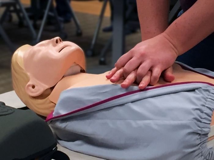
Heart disease patients and those suspected of having heart disease often face a battery of tests. Find out what you should expect if one of these noninvasive heart tests is in your future.
5:00 AM
Author |

This post is part of a mini blog series dedicated to bringing awareness about your heart health during the American Heart Association's national #HeartMonth. Miss the other stories? Catch up on 5 Eating Tips for a Healthy Heart, How To Check Your Blood Pressure at Home, How to Know If You're Having a Heart Attack and Getting Heart Healthy with a Mediterranean Diet.
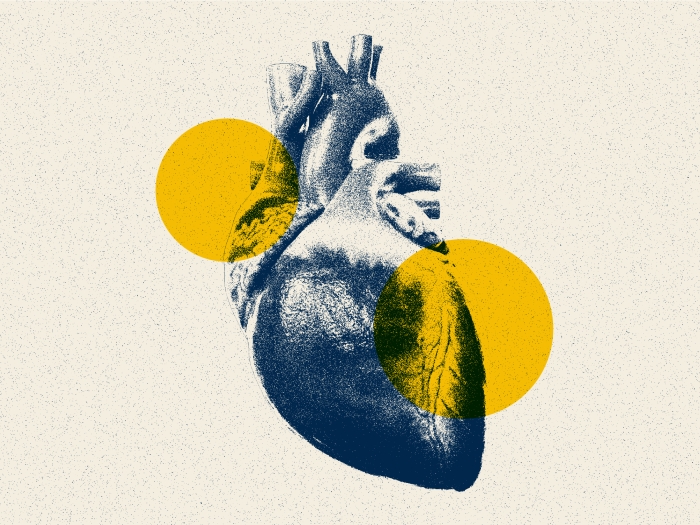
MRIs. CT scans. ECGs. There are a lot of test names you may hear when you visit your cardiologist, but what are all these tests and why do you need them?
If you have heart issues or are suspected of having a heart condition, your doctor may order an array of tests. But what's their purpose and what should you expect? Michigan Medicine cardiologist Venkatesh Murthy, M.D., offers a guide to the various noninvasive tests your doctor may order, including what they're designed to do.
1. Echocardiogram: Uses sound waves to produce images of your heart. This common test allows your physician to see how your heart is beating and how blood is moving through your heart. Images from an echocardiogram are used to identify various abnormalities in the heart muscle and valves. This test can be done while you're at rest or with exercise to elevate your heart rate (see exercise cardiac stress test below).
Reasons for the test:
-
Determine the cause of a heart murmur
-
Check the function of heart valves
-
Assess the overall function of the heart
2. Transesophageal echocardiography (TEE): Uses high-frequency sound waves (ultrasound) to make detailed pictures of your heart and the arteries that lead to and from it. The echo transducer that produces the sound waves for TEE is attached to a thin tube that passes through your mouth, down your throat and into your esophagus, which is very close to the upper chambers of the heart.
Reasons for the test:
-
Assess the function of heart valves
-
Follow heart valve disease
-
Look for blood clots inside the heart
3. Electrocardiogram (ECG or EKG): Measures the electrical activity of the heartbeat to provide two kinds of information. First, by measuring time intervals on the ECG, a doctor can determine how long the electrical wave takes to pass through your heart. Finding out how long a wave takes to travel from one part of the heart to the next shows if the electrical activity is normal, slow, fast or irregular.
Second, by measuring the amount of electrical activity passing through your heart muscle, a cardiologist may be able to find out if parts of the heart are too large or overworked.
Reasons for the test:
-
Monitor changes in heart rhythm
-
Determine whether a heart attack has occurred
-
Help predict if a heart attack is developing
4. Magnetic resonance imaging (MRI): Uses a magnetic field and radiofrequency waves to create detailed pictures of organs and structures inside your body. It can be used to examine your heart and blood vessels and to identify areas of the brain affected by stroke.
Reasons for the test:
-
Assess heart structure
-
Look for scar tissue within the heart muscle
-
Assess the function of heart valves
5. CT scan: An X-ray imaging technique that uses a computer to produce cross-sectional images of your heart. Also referred to as cardiac computed tomography, computerized axial tomography or CAT scan, it can be used to examine your heart and blood vessels for problems. It's also used to identify whether blood vessels in the brain have been affected by stroke.
Reasons for the test:
-
Assess the structure of the heart
-
Determine if blockages are present in the coronary arteries
6. Exercise cardiac stress test: Also called an exercise tolerance test (ETT), this test shows whether your heart's blood supply is sufficient and if your heart rhythm is normal during exercise on a treadmill or stationary bicycle. The test monitors your level of tiredness, heart rate, breathing, blood pressure and heart activity while exercising. This test may be done in combination with nuclear imaging or echocardiography.
MORE FROM MICHIGAN: Sign up for our weekly newsletter
Reasons for the test:
-
Determine the cause of chest pain, shortness of breath and weakness
-
Assess the health of the heart
-
Assess safety of exercise
-
Identify heart rhythm changes with activity
-
Find evidence of inadequate blood flow to the heart muscle during exercise
7. Pharmacologic stress test: Medication is given through an IV line in your arm to dilate the arteries, which increases your heart rate and blood flow, similar to the effects of exercise. This test may be done in combination with nuclear imaging, echocardiography or MRI.
Reasons for the test:
-
Determine the cause of chest pain, shortness of breath and weakness
-
Find evidence of inadequate blood flow to the heart muscle during exercise
-
Monitor or diagnose blockages in the coronary arteries
-
Assess risks for a heart attack
8. Tilt test: Often used to determine why you feel faint or lightheaded. During the test, you lie on a table that is slowly tilted upward. The test measures how your blood pressure and heart rate respond to the force of gravity. A nurse or technician keeps track of blood pressure and heart rate (pulse) to see how they change during the test.
Reasons for the test:
-
Assess dizziness or fainting spells
-
Identify heart rhythm changes
9. Ambulatory rhythm monitoring tests: Holter monitoring, event recorders and mobile cardiac telemetry (MCT) are ambulatory monitoring tests done to study your heart rhythm for a prolonged period of time on an outpatient basis.
Reason for the tests:
-
Look for evidence of heart rhythm problems that come and go and that are not apparent with a standard ECG
LISTEN UP: Add the Michigan Medicine News Break to your Alexa-enabled device, or subscribe to our daily updates on iTunes, Google Play and Stitcher.
10. Coronary angiogram: A type of X-ray used to examine the coronary arteries supplying blood to your heart. A catheter is inserted into a blood vessel in your arm or groin and fed up to your heart and coronary arteries. Special dye is then injected through the catheter and images are taken.
Reasons for the tests:
-
Identify narrowing or blockages in the coronary arteries
-
Evaluate pressures inside the heart

Explore a variety of healthcare news & stories by visiting the Health Lab home page for more articles.

Department of Communication at Michigan Medicine
Want top health & research news weekly? Sign up for Health Lab’s newsletters today!